What is the function of the optic cup?
The optic cup is used to diagnose glaucoma. The cellular strata that constitute the optic cup cover the entire cup margin. They also cover the lens at its front and stretch toward the pupil’s aperture. Each layer has a specific role in the formation of the retina.
What is the Cup in the optic disc?
The optic cup is the white, cup-like area in the center of the optic disc. The ratio of the size of the optic cup to the optic disc (cup-to-disc ratio, or C/D) is one measure used in the diagnosis of glaucoma. Different C/Ds can be measured horizontally or vertically in the same patient.
What is the function of the optic disc blind spot?
What is the purpose of a blind spot in the eye? The blind spot is where the optic nerve and blood vessels leave the eyeball. The optic nerve is connected to the brain. It carries images to the brain, where they’re processed.
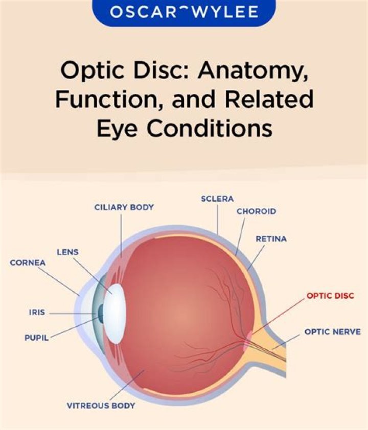
What is optic disc?
Optic disc: The circular area in the back of the inside of the eye where the optic nerve connects to the retina.
What is a cup eye?
1 : a small oval cup with a rim curved to fit the orbit of the eye used for applying liquid remedies to the eyes. 2 : optic cup. 3 : a usually rubber cup at the eyepiece of an optical instrument (such as binoculars) for keeping out extraneous light.
What causes cupping of the optic disc?
OPTIC DISC CUPPING. A hallmark of glaucoma is excavation or enlargement of the optic disc, referred to as cupping. The vast majority of pathologic cupping is caused by glaucoma. Disc cupping in the absence of elevated IOP may be caused by normal-tension glaucoma, which accounts for 20% to 30% of open-angle glaucoma.
What is the difference between optic disc and blind spot?
The optic disc or optic nerve head is the point of exit for ganglion cell axons leaving the eye. Because there are no rods or cones overlying the optic disc, it corresponds to a small blind spot in each eye. The optic disc is also the entry point for the major blood vessels that supply the retina.
Is optic disc same as optic nerve?
Optic disc: The circular area in the back of the inside of the eye where the optic nerve connects to the retina. Also called the optic nerve head.
How does the optic disc work?
The optic disc (optic nerve head) is the location where ganglion cell axons exit the eye to form the optic nerve. There are no light sensitive rods or cones to respond to a light stimulus at this point. This causes a break in the visual field called “the blind spot” or the “physiological blind spot”.
What is the function of optic nerve?
The optic nerve is a bundle of more than 1 million nerve fibers. Also known as the second cranial nerve or cranial nerve II (CNII), it is the second of several pairs of cranial nerves. It transmits sensory information for vision in the form of electrical impulses from the eye to the brain.
Does cup to disc ratio improve?
Clinical improvement in visual fields was correlated with the degree of improvement of cup:disc ratio (P = 0.025). Conclusion: Most patients showing a 40% lowering of IOP after glaucoma surgery show improved optic nerve morphology as measured by the HRT.
What is normal disc to Cup ratio?
The normal cup to disc ratio (the diameter of the cup divided by the diameter of the whole nerve head or disc) is about 1/3 or 0.3. There is some normal variation here, with some people having almost no cup (thus having 1/10 or 0.1), and others having 4/5ths or 0.8 as a cup to disc ratio.
What causes optic nerve cupping?
“Cupping” is the result of changes in the optic nerve related to optic nerve degeneration, where there is a backward bowing of the central part of the disc. When your optic disc is seen in three dimensions, the “cupping” can be very obvious to your eye doctor.
Is optic nerve cupping normal?
“Cupping”. The normal optic nerve has a healthy appearing “rim” of tissue, which is assessed by both the contour of the rim as well as the color. “Cupping” is the result of changes in the optic nerve related to optic nerve degeneration, where there is a backward bowing of the central part of the disc.
Does large cupping of optic nerve mean glaucoma?
The second most significant risk factor for the development of chronic open-angle glaucoma is the size of the central cup “cupping” of the optic nerve head. The cupping of the optic nerve means the size of the depression in the middle of the nerve when viewed from the front of the eye. When there is damage to the optic nerve, the cupping increases.