What is the LGN for?
In addition to retinal afferents, the LGN receives input from multiple sources including striate cortex, the thalamic reticular nucleus (TRN), and the brainstem. The LGN therefore represents the first stage in the visual pathway at which cortical top-down feedback signals could affect information processing.
Where is the LGN?
They wrap around the midbrain and cross the medial surface of the temporal lobe, and 80% of them then terminate in a synaptic relay called the lateral geniculate nucleus (LGN), located in the dorsal part of the thalamus. The LGN is thus the major target for each optic tract.
How does the LGN work?
The LGN brings retinotopic maps from both eyes into register to make it easy for cortex to combine inputs from the two eyes. Only 10% of inputs to LGN come from the retina. 90% are modulatory inputs from cortex and the brainstem.
What does LGN stand for psychology?
The lateral geniculate nucleus (LGN) (lateral geniculate body or lateral geniculate complex), located in the thalamus, functions as a relay center for the visual pathway and receives the majority of its sensory input from the retina of the eye.
What is Retinotopic mapping?
Retinotopy (from Greek τόπος, place) is the mapping of visual input from the retina to neurons, particularly those neurons within the visual stream. Even more complex maps exist in the third and fourth visual areas V3 and V4, and in the dorsomedial area (V6).
What happens if the LGN is damaged?
In humans and other primates, visual information is transmitted from the retina to a part of the brain called the lateral geniculate nucleus (LGN), before reaching the primary visual cortex (V1). If the V1 is damaged, conscious vision is lost in the area of the visual field that corresponds to the damage.
Do the layers of the LGN separate left eye and right eye inputs?
Ipsilateral and contralateral layers Both the LGN in the right hemisphere and the LGN in the left hemisphere receive input from each eye. However, each LGN only receives information from one half of the visual field.
What is V2 in the brain?
V2. Visual area V2, or secondary visual cortex, also called prestriate cortex, is the second major area in the visual cortex, and the first region within the visual association area. It receives strong feedforward connections from V1 (direct and via the pulvinar) and sends strong connections to V3, V4, and V5.
Is V1 Retinotopic?
Area V1 has retinotopic organization, meaning that it contains a complete [map of the visual field | visual map] covered by the two eyes. For instance, 50% of the area of human V1 is devoted to the central 2% of the visual field (Wandell, 1995).
Does retina have Retinotopic mapping?
What causes Akinetopsia?
Several causes have been described to cause akinetopsia. These include infarction, traumatic brain injury, neurodegenerative disease such as Alzheimer’s ( visual variant of Alzheimer’s disease/ posterior cortical atrophy), epilepsy, hallucinogen persistent perception disorder (HPPD), and medication adverse effect.
Can damage to the visual cortex cause blindness?
Cortical blindness is an important cause of blindness due to damage to the occipital cortex. It is commonly associated with posterior circulation stroke. Hence recognizing it can lead to proper management and improved outcome.
Does LGN receive input from both eyes?
Both the LGN in the right hemisphere and the LGN in the left hemisphere receive input from each eye. However, each LGN only receives information from one half of the visual field.
What part of the brain controls vision?
occipital lobe
The occipital lobe is the back part of the brain that is involved with vision.
What part of the brain controls color vision?
fusiform gyrus
B&W stimuli (for both objects and non-objects), confirming that the fusiform gyrus is the brain center for color perception.
What does a person with akinetopsia see?
Akinetopsia (Greek: a for “without”, kine for “to move” and opsia for “seeing”), also known as cerebral akinetopsia or motion blindness, is a rare neuropsychological disorder in which a patient cannot perceive motion in their visual field, despite being able to see stationary objects without issue.
Why am I seeing things in slow motion?
This phenomenon is known as akinetopsia, the loss of motion perception. Patients do see the objects but cannot perceive their movement for some time. The so-called Zeitruffer phenomenon is similar to akinetopsia and manifests itself as an altered (usually slowed down) perception of the velocity of the moving objects.
Is CVI permanent?
Here’s what we know: While Cortical Visual Impairment/Cerebral Visual Impairment (CVI) can’t be completely cured, appropriate educational supports might help. In fact, plenty can be done with the hope of improving your child’s vision. Here are some key things to keep in mind: CVI can evolve over time.
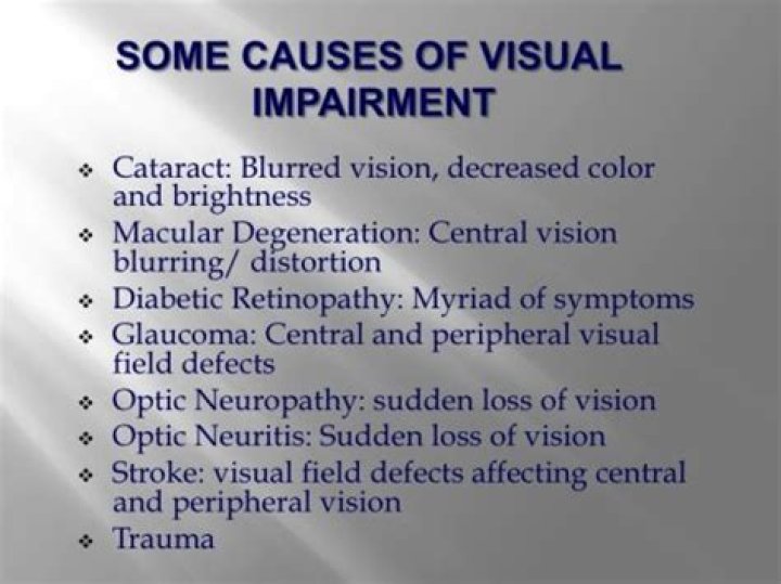
Is a visual impairment a disability?
If a consultant ophthalmologist has registered an individual as blind or partially sighted, then they will automatically meet the definition of a disabled person under the Equality Act (2010).
Is eye part of brain?
The eye is the only part of the brain that can be seen directly – this happens when the optician uses an ophthalmoscope and shines a bright light into your eye as part of an eye examination. And if pressure in the brain increases, perhaps due to a brain tumour, we can see this as a swelling of the optic nerve.