What happens microglial activation?
The chronic activation of microglia may in turn cause neuronal damage through the release of potentially cytotoxic molecules such as proinflammatory cytokines, reactive oxygen intermediates, proteinases and complement proteins.
How do microglia become activated?
Microglia become activated following exposure to pathogen-associated molecular patterns (PAMPs) and/or endogenous damage-associated molecular patterns (DAMPs), and removal of the immune-suppressive signals. Activated microglia can acquire different phenotypes depending on cues in its surrounding environment.
What is the role of microglia in the brain?
Microglia are the primary immune cells of the central nervous system, similar to peripheral macrophages. They respond to pathogens and injury by changing morphology and migrating to the site of infection/injury, where they destroy pathogens and remove damaged cells.
How do microglia get past the blood brain barrier?
We demonstrated that microglia respond to inflammation by migrating towards and accumulating around cerebral vessels, and that this begins before any detectable change in BBB permeability. Surprisingly, our data suggest that the initial microglial contact with cerebral blood vessels actually protect BBB integrity.
Is microglia part of a neuron?
microglia, type of neuronal support cell (neuroglia) occurring in the central nervous system of invertebrates and vertebrates that functions primarily as an immune cell.
How do you stop microglial activation?
Resveratrol has been shown to inhibit the activation of microglia and reduce the production of pro-inflammatory factors through intracellular cascades of signaling pathways such as MAPKs, phosphoinositide3-kinase (PI3-K)/Akt, and glycogen synthase kinase-3β (GSK-3β) pathways.
Do activated microglia proliferate?
Microglia are resident brain macrophages that become activated and proliferate following brain damage or stimulation by immune mediators, such as IL-1β or TNF-α. In conclusion, these findings indicate that microglial proliferation in response to IL-1β or TNF-α is mediated by hydrogen peroxide from NADPH oxidase.
Do microglia contribute to the blood-brain barrier?
Current data indicate that M1 pro-inflammatory microglia contribute to BBB dysfunction and vascular “leak”, while M2 anti-inflammatory microglia play a protective role at the BBB.
Does microglial form blood-brain barrier?
The blood-brain barrier (BBB), constituted by an extensive network of endothelial cells (ECs) together with neurons and glial cells, including microglia, forms the neurovascular unit (NVU). The crosstalk between these cells guarantees a proper environment for brain function.
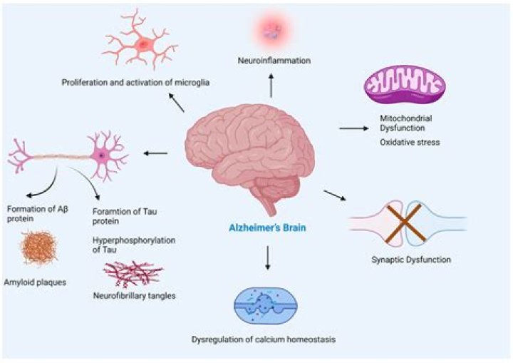
What is the role of microglia in neurodegenerative diseases?
We will review the role of microglia in neurodegenerative diseases such as Parkinson’s disease, Alzheimer’s disease and frontotemporal dementia (FTD), where microglial activation stands as one of the key components linked to the progression of the pathology and the symptoms severity.
What is microglial activation and how is it regulated?
Microglial activation is involved in the progression of different neurodegenerative diseases. Microglial phenotype regulation is mostly dependent on their interaction with molecules released by surrounding cells (neurons, microglial cells, astrocytes, etc.) through membrane-bound pattern recognition receptors (PRRs).
What are microglia sensitive to?
Microglia are sensitive to many stimuli or changes in the microenvironment of the CNS.
How many microglia are there in the human brain?
Depending on the anatomical region, microglia account for 0.5–16.6% of the total cell population in the human brain ( Lawson et al., 1992) and 5–12% in the mouse brain ( Mittelbronn et al., 2001 ).